


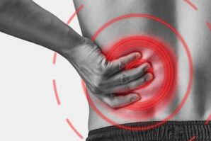
Lumbar osteochondrosis is a chronic degenerative-degenerative disease of the lumbar spine that affects the structure of intervertebral discs and several localized lumbar vertebrae.It affects people with a predominantly working age.It manifests itself in various symptoms, the main one of which is pain in the lower back and legs, limiting movements in the lower back.For diagnosis, research methods such as radiography, computed tomography or magnetic resonance tomography of the lumbar spine are used.In this article, you can more detail with the causes, symptoms and methods to diagnose lumbar spine osteochondrosis.
Osteochondrosis is the result of body aging.These or other signs of this disease can be found in almost everyone (!), From 25 years.But here is the severity of these changes, the rate of their progression, the degree of clinical manifestations depends on many causes, especially about how a healthy lifestyle leads a specific person.Moderate physical activity, mandatory morning gymnastics, the right pose of the body when performing a work number (garden, construction, banal cleaning of the house and so on), the orthopedic mattress is those that prevent the development of lumbar spine osteochondrosis.
According to statistics, spine osteochondrosis in 80% of cases is the cause of back pain.
How does osteochondrosis develop?
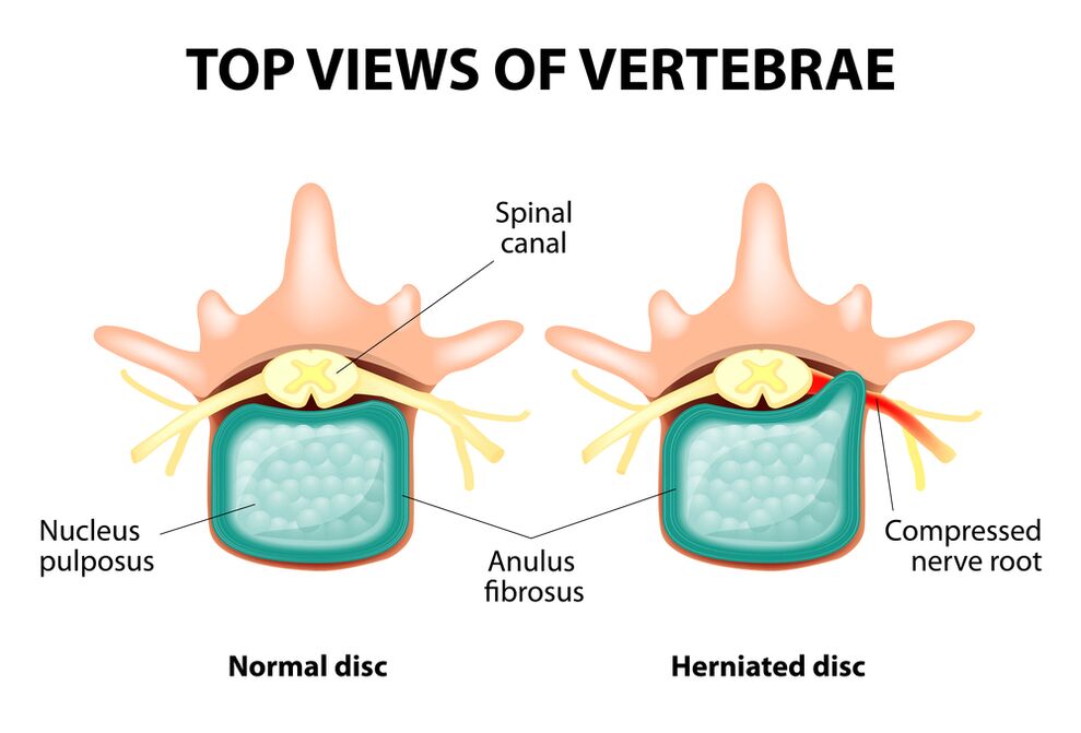
The whole column consists of separate vertebrae, among the bodies of which there are intervertebral discs.That is, between the two vertebrae is a disc.The disc consists of a gelatinous (pulpitus) nucleus and a fibrous ring.The core contains a lot of water and provides depreciation and flexibility of the spine.The fibrous ring is located along the outskirts of the jacket core, as if holding it inside.
With a prolonged increased load in the jetty core, it changes its physiological properties, loses water and dry and eventually sequences: the disc is flat and the vertebral bodies approach.Along with these processes, in the core jacket, the fibrous ring loses its elasticity and, under the influence of mechanical loads, begins to project itself.This is called protrusion.Then the fibrous cracks and a gelatinous core falls through the resulting gaps: a hernia of the disc occurs.A graph of two adjacent vertebrae and a record located between them, called the spill segment, acquires excess mobility, thus increasing the load in the nearby segments.The overload of neighboring segments triggers a similar pathological process in them.These changes are called osteochondrosis.
To somehow ensure the stability of the spine, bone growths are formed along the edges of the vertebral bodies, increasing the support area.This phenomenon is called spondylosis.Changes in the joints between the vertebrae are called spondylo arthrosis.Generally, all three pathologies - osteochondrosis, spondylosis, spondil arthrosis - walk nearby.
Reasons
Why does osteochondrosis occur?To date, there are several theories of the occurrence:
- Mechanical Theory: Perhaps the main reason should be considered a regular increased load in the spine.This is why osteochondrosis is an almost mandatory destination for engines, miners, builders and people of such professions.The occurrence of osteochondrosis in the lower back is mainly associated with the inclinations and survey of gravity, forced an uncomfortable pose of work;
- Another factor in development is the incorrect posture, sitting in the wrong pose, which is especially relevant to mental workers;
- Sometimes the role is played by the hereditary characteristics of the spine structure and the nutrition of its individual structures;
- Traumatic theory: any trauma in the spine (even the most insignificant) is able to launch a degenerative process;
- Hormonal metabolic disorders and endocrine diseases may adversely affect metabolism in spinal spine tissues and contribute to the development of osteochondrosis;
- The theory of age implies the natural wear of the discs in the life process.
Rarely, only one of these theories can explain the occurrence of osteochondrosis in each case.More often, at the same time, several factors are "guilty."
In the occurrence of lumbar spine osteochondrosis, overweight plays an important role as it is an overload for the spine of the spine.The higher the body mass index (degree of obesity), the more changes the more pronounced in the spine are usually.Among other reasons, causing the appearance of osteochondrosis, it can be observed:
- sedentary lifestyle;
- Imorly Nutrition (Fast Food, Excess Sweet and Semi -Acabados: All of this leads to an imbalance of traces) and lack of fluid;
- Anomalies of the column structure (for example, the presence of an additional lumbar vertebra);
- constant use of high shoes;
- Pregnancy (due to excess load in the lumbar spine);
- Sudden termination of training in people professionally involved in the sport;
- Smoking and alcohol abuse: as factors that accelerate the aging process in the body.
Symptoms
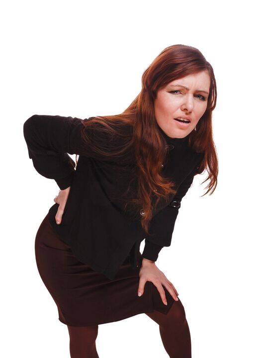
The main manifestation of lumbar spine osteochondrosis is pain.The nature of pain, the place of occurrence and the direction of distribution depend on which receptors are irritated, that is, as approximate changes on the disc and surrounding tissues, there is protrusion or hernia, in which direction of protrusion was formed and so on.
Reflex and compression syndromes are distinguished with lumbar spine osteochondrosis.
Reflex syndromes develop in cases where the affected disk fibrous ring receptors, ligaments and joint capsules located nearby are irritated.They are reflective because, besides pain, they are accompanied by muscle-so-called, vegetative-tasular or neurodistrophic changes, ie reflex irritation is transmitted to other structures, causing symptoms mainly from the soft tissue side.
Compression syndromes occur as a result of compression (compression) of nerve roots, blood vessels or spinal cord formed by osteochondrosis by changes.

Syndromes Reflex of the lumbar spine
Lumbago(Feeling): acute sudden pain in the lower back, which occurs with a clumsy movement or at the time of physical tension (much less frequency - without apparent reason).It is believed that the occurrence of lumbago is associated with the movement of a jacket nucleus within the fibrous ring, ie it develops in the early stages of osteochondrosis.Often the pain is described as "feeling", "the stake was stuck in the lower back."Patients freeze in the pose where pain took them.Lower movement causes an increase in pain (sneezing, coughing, an attempt to turn into bed, move the foot).If a person was in a sloping position at the time of lumbago development (which happens more often), he cannot straighten himself.A muscle tension pronounced in the lumbar spine occurs reflexively.Throughout the vertebrae in this area, a muscle roll is felt, which is sometimes visible to the naked eye without touch, and muscle tension is so pronounced.Feeling painful for the patient.Increased muscle tone plays an immobilizing role, protecting the affected lumbar segment from pathological mobility, which can cause deterioration in the state.The natural curve of the spine of the spine in the lower back (lordosis) is flat, perhaps the curvature (scoliosis) is possible due to muscle tension.
Lumbalgia- Another lumbar level reflex syndrome.This term also means the presence of pain in the lower back.But unlike lumbago, the pain does not arise sharply, but gradually, within a few hours or even days.The pain is stupid, the moderate intensity, intensifies during the movements, in a sitting position or standing when moving from one position to another.A little relief brings the position of lying down or back with a roll in the lower back, but the passive increase in the straightened leg in this position causes increased pain in the lower back (Lassa symptom).Palpation of the lumbar spine is painful, but the reflex tension of the muscles is less pronounced than with lumbago and sometimes absent.Lumbar spine movements are limited but possible.This means that the patient can bend and to the sides for a certain level (and then the pain intensifies).
Sciatica- Another variety of lumbar level reflex syndrome.By this term, it means pain in the lower back, which gives the buttock and leg (on the rear surface).The pain is different, especially painful, but can intensify periodically by the type of "fireplace" in the leg.As with lumbalgia, it intensifies with any movements, walking, stressing, decreases in the back.Lassa's symptom is usually positive.Palpation of the lumbar spine is painful, in addition to pressing some points (for example, in the middle of the line that separates the buttock from the thigh, in the middle of the back of the thigh, in the middle of the popliteal fossa).There is tension of the lumbar muscles.In front and side inclinations are limited.
Lumbar spine compression syndromes
The clinical feature depends on which structure is subject to compression.
Among the vertebrae in each intervertebral hole are the nerve roots (spinal nerves): left and right.If pathological formations for lumbar spine osteochondrosis (mainly disc discs) squeeze the roots, then radiculopathy develops, whose symptoms differ for each root.Common to all radiculopathies in the lower back is increased pain during sneezing, cough, movement in the lower back (especially leaning forward), the presence of muscle tension in the lower back, restriction of movements in the lumbar spine.The following types of lumbar spine radiculopathies are more common:
- Radiculopathy L1, L2, L3: Pain occurs in the lower back, give it to the early thigh.In the same area, paresthesia is possible (a sensation of tracking goose bumps, numbness), superficial sensitivity is disturbed (an acute touch of the usual is not distinguished, the feeling of cold and hot) is lost.The reflex of the knee decreases, the weakness of the thigh quadriceps is revealed;
- L4 Radiculopathy: The pain of the lower back gives the front -line of the thigh, the inner surface of the knee joint and slightly lower along the inner surface of the leg.In the same areas, paresthesia is felt and surface sensitivity is lost (reduced).Weakness in the quadriceps thigh muscle also develops, the reflection of the knee decreases;
- L5 Radiculopathy: One of the frequent locations.The pain gives the buttock along the outer edge of the thigh along the front surface of the leg to the inner edge of the foot and thumb.Paresthesia is felt here, superficial sensitivity is disturbed and a pain is given here when sneezing and coughing.In addition, there is a difficulty in extending the thumb of the foot, since the muscle that performs this action is innervated by the Kine L5.Sometimes it is difficult to stand with an exposed foot;
- Radiculopathy S1 is also often found with lumbar spine osteochondrosis.The pain gives the buttock along the outer edge of the thigh along the outer edge of the leg to the outer edge of the foot and the 5th finger, jumps.These zones are characterized by a feeling of paraesthesia, a decrease in surface sensitivity.The reflection of Achilles is reduced.With damage to this column, the weakness of the leg muscles and the foot flexors develops, so that it develops and walking in the socks is difficult.
Simultaneous development of radiculopathies of various roots is possible, this is especially characteristic of L5, S1.It turns out that a hernia tightens several roots.
If the disk herd is back, you can tighten the spinal cord.This is only possible when the hernia is located at the upper reference point, as there are no spinal cord vertebrae below the Lumbar II vertebra (the spinal cord roots are subjected to compression and the horse's tail syndrome develops).
If the back vessels are subjected to squeezing, which perform blood flow to the spinal cord, then, in the case of an acute circulatory disorder, a spinal course develops and with prolonged compression - myelopathy.Myelopathy is manifested by bilateral weakness of the leg muscles, starting with the foot and gradually progressing.The sensitivity in the legs is disturbed, the reflection of Achilles is lost and then the knee.It is possible to emerge disturbances of urination (frequent and "imperative" desire, requiring immediate satisfaction, urinary incontinence).
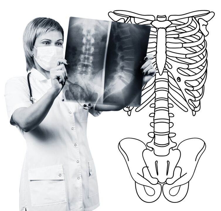
Diagnostic methods
The diagnosis of lumbar spine osteochondrosis is based on clinical data and additional research method data.The main function belongs to methods such as:
- lumbar spine radiography;
- computed tomography of the lumbar spine;
- Magnetic resonance tomography of the lumbar spine.
Lumbar spine radiography is necessarily performed in 2 projections mutually perpendicular to it and the straight side.Such images allow you to see the shape, contours and structure of vertebral bodies, the height and shape of intervertebral discs, the abnormalities of the spine and natural curves.To display intervertebral joints and intervertebral holes, radiograms are produced in oblique projections.To identify the pathological mobility of individual lumbar segments (which is a sign of osteochondrosis), the radiography is performed under functional study conditions, ie, flexion and extension of the spine.Normally, you can clearly see the height change in the intervertebral discs in the front or rear sections according to the direction of the body tilt, with osteochondrosis due to the functional block of one of the segments, the height of the disk does not change when double or stress.With pathological mobility, the displacement of the vertebrae is determined back or forth.The main x -ray signs of osteochondrosis include narrowing of intervertebral slit, pathological mobility and vertebral bodies, deposition of salts in disc tissue (calcification), the formation of regional vertebral bodies, compaction of vertebrae on the border with the affected disc (accumulated the radiography of the lumbar column is a research routine, which gradually that gradually that gradually that gradually that gradually that graduallyIt loses its meaning in relation to the antecedents of the active implementation of new and more informative research methods (CT and RM). The lumbar department radiography is now used as a method of diagnosing screening.
Lumbar column CT is also performed using X -Ray radiation, but the radial load on the body is much smaller than with the X -ray. The study is performed on the table of a special device - a computer tomography is absolutely painless.The resulting images are processed using a computer and allow you to significantly see more structures than with the column radiography.
Magnetic resonance imaging is a method in which electromagnetic radiation is used to create images.The study is also conducted in the position on the table, which calls the Tomography Chamber.Magnetic resonance imaging is harmless and painless.
CT or magnetic resonance imaging of the lumbar spine allows you to see all the structures of the spine, carefully examine the intervertebral discs (and the jacket and the fibrous ring) and the intervertebral holes, the contents of the spinal canal.Even a slight protrusion of the intervertebral disc will not go unnoticed.These methods (especially magnetic resonance imaging) allow to determine the direction of the herniated disc, if any, the degree of compression of the nerve roots, the spinal cord.Thus, these research methods are much more informative in the diagnosis of lumbar spine osteochondrosis than radiography.In addition, they allow not only osteochondrosis, but also other diseases (tumors, circulatory disturbances in the spinal cord, abscesses, congenital defects of the spine structure and spinal cord), which is important during the differential diagnosis of back pain causes.
Lumbar spine osteochondrosis is a disease that most often causes back pain.It is, in fact, the destruction of intervertebral discs.Due to lumbar spine osteochondrosis, a person usually loses working capacity, because, besides pain, the disease can lead to a violation of spine mobility, the inability to sit, stand and walk.Symptoms of this disease are non -specific and require additional research methods to accurately confirm the diagnosis.The most informative and safe of modern osteochondrosis diagnostic methods is the magnetic resonance imaging of the spine.